Macular Pucker
Your macula is a small area in the center of your retina. This small center area allows you to see fine details clearly. A
normal macula should remain flat against the back of the eye. Macular pucker and or epiretinal membrane is a condition in
which scar tissue develops on the macula (central part of vision). There is a varying degree of severity with a macular
pucker. Macular puckers or epiretinal membranes should be closely monitored by your retinal specialist. Your retinal
specialist will be able to monitor the progression of the scar tissue. Some macular puckers require treatment to regain
visual acuity and or stop the progression of visual loss.
The vitreous (clear gel substance that fills your eye) over time will begin to shrink and pull away from the retina. This is a
normal process that comes with age. As the vitreous pulls away, scar tissue may develop on the macula. The scar tissue
may wrinkle the retina causing a visual disturbance.
With a macular pucker you may notice your vision is blurry centrally. You may also notice, when looking at a straight
line, that it has a bend or wave in it. Words may appear crooked when reading a book or paper and it may become
increasingly difficult to enjoy your favorite hobbies over time.
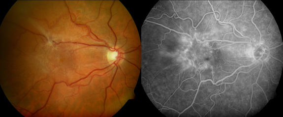
Your doctor can diagnose by doing a dilated exam, as well as fluorescein angiogram and/or ocular coherence
tomography. Fluorescein angiogram and ocular coherence tomography are both important test that are done to help
diagnose this condition.
Treatment for macular pucker is not always required or suggested by your doctor. Some macular puckers are mild and
do not worsen. For more severe macular puckers, surgery can be performed by your retina specialist. You and your retina
specialist can decide if surgery is warranted to help regain visual acuity or stop the progression of visual loss or
distortion.
Routine dilated eye exams are important in detecting a macular pucker early in its progression.